This blog entry has long been in the making. I started writing it in April 2014. The topic of
this post will be bars, and whether or not they can “circle around the frog”,
“smear in the sole” or “overlay the sole in a thin veil”.
Bars migrating or smearing or pushing up in the capsule is a
central theme in the ABC hoof print trim. According to the inventor of the ABC
trim, Cheryl Henderson, many pathologies are attributed to bars having migrated
to places they should not be, or getting imbedded in sole. Often times,
painting a picture where bars overlay the sole and crush new sole tubules
underneath lead beginner trimmers (including me in early 2011) to dig out supposed
bar material, in search for some sole underneath, but instead finding themselves
having “quicked” (i.e. hit blood) their horses (thankfully excluding me!).
I personally started questioning this idea, that bar smears
or migrates when I was removing it over the course of 6 months to one year,
only to find, that my horses a) became extremely sensitive on their soles and
b) the supposed “bars” growing back every time exactly the same way as I found
them 4-6 weeks prior. So I
concluded that something must not be correct with this theory.
Cheryl Henderson believes that her own research has proven that
bar (or better the “zona alba” of the bar, i.e. the inner unpigmented wall of
the bar) can migrate and circle around the frog. However, what people without any
formal research training may not realize, this
research is merely an observation combined with a hypothesis and not a proof whatsoever.
The scientific process usually has several steps:
1)
Most research starts out with an observation.
2)
Next, one proposes a mechanism for that observation.
3)
Third, one proves
that this mechanism is indeed able to generate the observation. The proof is
NOT the observation itself, or otherwise it would be an entirely circular
argument.
4)
One states exceptions to the rule, or how significant the observation is. For
example, of all cadaver hooves that were analyzed, the patterns of bar
circling/embedding around the frog has only ever been observed on white hooves.
Not on black hooves. (Note: on white hooves bar and sole are basically
indistinguishable, whereas on black hooves white bar circling around or
embedding into sole should easily be distinguished from black sole).
5)
One submits the research to a scientific peer-review process stating truthfully
all observations that have been obtained, and not only the ones that fit the
mechanism proposed by the author
6)
The scientific community then decides if a study
is worthwhile publishing provided all rules
of good scientific practice have been followed.
In the particular case of Cheryl Henderson’s beetle cadaver study, if it
would ever have reached step 5), the reviewers should have severely questioned
the results based on point 4). It is
HIGHLY unlikely that a mechanism of bar circling around the frog should be
limited to white hooves only.
So here is now a more detailed description of the research.
It is pretty simple, but I admit, very cool and also very appealing to people
who do not know any better: she throws a hoof in a container with dermestid
beetles. These beetles are used by taxidermists (and crazy biologists at times)
to clean bones from flesh. An example of one hoof after
being exposed to the beetles is shown below. This picture has been circling Facebook for years now:
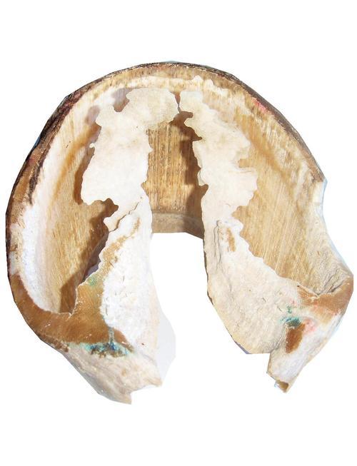
What remains of the hoof after the beetles have spent some time with it is the wall,
the bars, and some material that is attached to the bars but have a flat
appearance encircling the frog (which of course is not there anymore, as the
bugs most likely ate that one first, as it is about the softest material on the whole hoof). Her conclusion is that the latter, since
it is connected to the bar, must have grown from the bar, and therefore
represents bar. Then she goes on explaining that very many horses have bar
circling around the frog and this causes them major damage.
My criticism would be that there is no proper scientific protocol being followed here.
It is just an observation, followed by a hypothesis. And that is fair enough. This is the first step in all research.
The problem lies in the fact that Cheryl considers this observation as proof. It is a good observation, but the conclusions and implications she insists it
is showing do not exist. There is also no cautioning that there could be other
explanations for the patterns observed, which, in science, until something is
really proven several times independently, is almost always the case. Nor any
mechanism that would explain how bar can so far outgrow its corium.
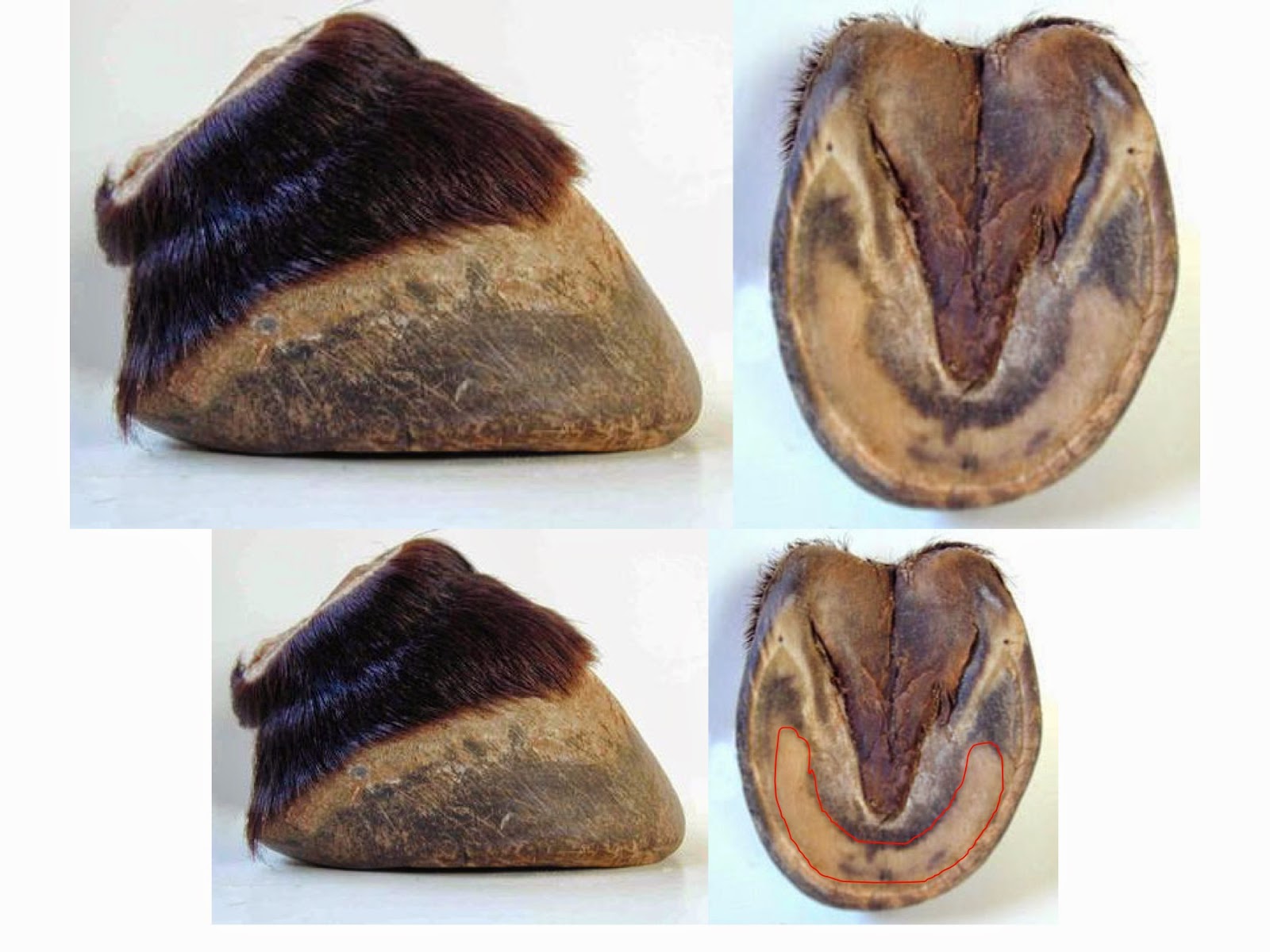
This is a cadaver hoof. I need to point out that this is NOT the same hoof as pictured above. Cheryl Henderson does not provide any pictures of the hoof depicted above before the beetles started to work on it. But the hoof below has
the typical "bars circling around the frog" issue, that Cheryl is so
particular about. I have circled the supposed "bar" in black.
This is the other side of that
same hoof with the coffin bone sitting in the hoof capsule, but the navicular bone missing.
The blue arrows point to the true end of the bar, where the lamellae of
the white line end. What is forward of the end of the bar are straight horn
tubules, that look a lot like the ones from the hoof wall. They just somehow
look different than normal sole tubules, somehow stronger and thicker. Yet, in
my opinion, this is not bar that "smeared" or "migrated",
but sole, maybe of some slightly different anatomy as sole further out on the
hoof, i.e. not right next to the frog and bar. After all, this sole finds
itself at a “junction” the junction between frog and sole and bar. It has to
connect the bars with the sole and the frog. It must be a rather difficult task and one of massive importance. Junctions usually are weak points,
so it would make total sense if that junction would be somehow re-enforced by
having some stronger sole tubules.
I am not saying that my
interpretation is necessarily correct. It is just as much of a hypothesis as
Cheryl’s interpretation is one, that this material is inner bar wall smeared out from the back of the hoof. Future research would need to look in the detailed
anatomy of hooves and in particular that junction between bar, sole and frog on
many hooves, black, white, from lots of different living environments etc.
Now, this bar/sole/frog junction
horn is exactly the horn that the bugs don't like eating.
As I said above, in my opinion, the fact that the beetles don't like eating this structure does not prove it is bar. It is attached
to bar, but this must not mean that is indeed bar. It is more likely that the sole near the bar-sole-frog
junction is anatomically somewhat different. And this in turn would mean that the
concept of trying to dig out bar around the frog is misleading. Yes, this cadaver
hoof has some accumulation of excess material around the frog, I don't think anybody denies that, but it does not need to be
dug out in search for sole "underneath". It simply needs to be
smoothed off to the level of exfoliated sole around it, and that's it.
Now comes my evidence (still no proof!), that the material that circles around the frog is not bar, but compressed sole callus, that, on white hooves, can be difficult to distinguish from bar.
Here is one of my geldings hooves from April 14th of this year,
when I started to work on this blog post. Short background on him is on top of
the slide. If I would have approached Cheryl for help with this hoof (which I
did 3.5 years ago) she would have told me to lower his heels, his bars and
remove the material that I have marked in red in the following slide, claiming
it would be bar that smeared out and overlaying the sole, crushing it underneath. She would have told me to take sliver after
sliver and inspect it under the microscope so that I would know if it is bar or
sole (but no information is given that this is even possible to distinguish under the microscope). It is a white hoof, of course, so bar and sole are hard to distinguish
(to the untrained eye, for me it is simple now, as I know those hooves in and
out).
This is the area (circled in red) that I would have been told to remove, representing bar smear:
 I have trimmed my horse for more
than 3.5 years now, in a rather strict 4 week schedule. For the last year or so
I have help by an AANHCP, who checks on my work periodically. I know with 100% certainty that this
material is not bar. I have defined the bars 100ds of times, all three layers
of it, pigmented and unpigmented wall and white line. If one looks carefully
one can see the white line in the bars straight and not laid over.
I have trimmed my horse for more
than 3.5 years now, in a rather strict 4 week schedule. For the last year or so
I have help by an AANHCP, who checks on my work periodically. I know with 100% certainty that this
material is not bar. I have defined the bars 100ds of times, all three layers
of it, pigmented and unpigmented wall and white line. If one looks carefully
one can see the white line in the bars straight and not laid over.
In this slide I have marked the
rough position of the coffin bone. It is quite clear in my mind that this red
line tracks pretty well the coffin bone above.
And again here, in comparison to
the material that the bugs don't like eating.

To me, all the evidence taken
together points to a model where sole around the frog can take on various
consistencies, callous and become really hard and sometimes look shiny and slick, so that beetles abstain from
eating it, at least as long as they have anything else to eat. This is because
the coffin bone exerts so much pressure, and for my horses, who live on a sand
dune, the sand (or snow) packs in their hooves and creates a lot of counter pressure. The
sole adapts to those pressures and callouses quickly and massively to provide the hoof with adequate
protection. Does it look necessarily pretty and beautiful, like the mustang feet? No! Absolutely not, but these feet could master ice and snow without one slip and one ouch and that is what counts. Mustangs don't spend 3 months on 3 feet of snow. At least not the ones we take as models these days (the Australian brumbies or the Great Basin mustangs). Hooves do look different in every single environment, and that makes them so special, their adaptability.
In my mind, there is undoubtedly
need for more research to study the different appearances that sole can take on
in different environments, and also the anatomy of that bar-sole-frog junction.
Linda Cowles has made a nice effort with respect to sole appearances. I have referred to her blog before:
http://mollyshoofjourney.blogspot.com/2014/08/the-different-appearances-of-sole-very.html
Clearly, noone has all the answers yet, and we all have to stay open minded and try and learn what it is the horse tries to tell us.